Exploring the Quorum Quenching Properties of Premna serratifolia on Pseudomonas aeruginosa
In-Silico & In-Vitro Antimicrobial Resistance
| Year | 2025 |
|---|---|
| Location | PES University |
| Tags | Docking, GROMACS, MMPBSA, Biofilm assays |
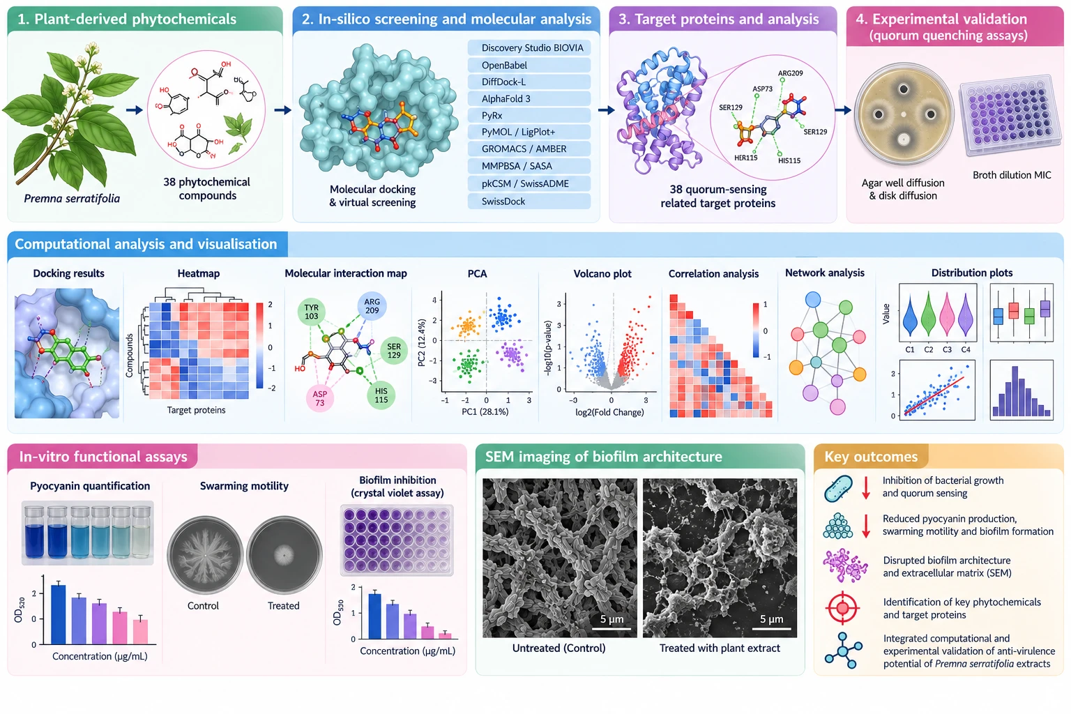
Wet-lab and computational study of quorum quenching: docking and MD of 38 phytochemicals against 38 quorum-sensing targets, plus MIC, pyocyanin, swarming and biofilm assays and SEM imaging of disrupted biofilms.
Aug 2023 – Dec 2025 at PES University, supervised by Dr. Reshma S. V..
- 01Phytochemical library
Assembled 38 phytochemicals from Premna serratifolia and 38 quorum-sensing-related target proteins of Pseudomonas aeruginosa, with structures prepared and predicted where needed.
AlphaFold 3, OpenBabel, Chimera, PyMOL - 02Docking & virtual screening
Screened every compound against every target to find the strongest binders, then mapped the protein–ligand interactions behind each hit.
Discovery Studio BIOVIA, PyRx, DiffDock-L, SwissDock, LigPlot+ - 03Molecular dynamics
Simulated the top complexes to check they stay bound over time, and estimated binding free energies and surface exposure.
GROMACS, AMBER, MMPBSA, SASA - 04Drug-likeness & data analysis
Checked ADMET and drug-likeness, then summarised the screen with heatmaps, PCA, correlation, clustering, network and violin plots.
SwissADME, pkCSM, Python plotting - 05Wet-lab assays
Tested plant extracts at the bench for antibacterial, quorum-sensing and anti-virulence effects: agar well and disk diffusion, broth-dilution MIC, pyocyanin quantification, swarming motility and crystal-violet biofilm inhibition.
Agar well diffusion, Disk diffusion, Broth dilution MIC, Pyocyanin assay, Swarming assay, Crystal violet - 06SEM imaging
Imaged biofilms before and after extract treatment to see the structural disruption and reduced extracellular matrix.
SEM, ImageJ